Hemispheric Disparities in Neurodegeneration
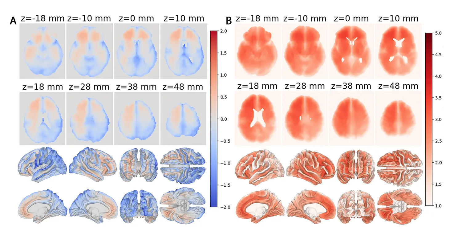
Researchers have unveiled a novel artificial intelligence tool that maps brain aging through detailed MRI analysis. Published in the Proceedings of the National Academy of Sciences, this study demonstrates that biological aging does not occur uniformly across the brain. The findings highlight significant disparities between the left and right hemispheres, offering new insights into neurodegenerative processes.
Wellness insights
 Brain Wiring Explains Why Trauma Victims Struggle To Find Joy
 Healing Journey: A Therapist Reflects on Processing Deep Childhood Trauma
 Forest walks boost mood through sound and biodiversity awareness
 What Counselling in Two Different Countries Taught Me: Hansel Wong on Connection, Curiosity, and Culture
The team developed an advanced algorithm capable of processing high-resolution magnetic resonance imaging data. This technology allows for a granular examination of specific brain regions rather than treating the organ as a single unit. By applying this method to large datasets, scientists identified distinct patterns of tissue degradation. The results suggest that the right hemisphere often exhibits markers of accelerated biological age compared to its counterpart on the left.
The uneven distribution of aging signs has profound implications for understanding Alzheimer’s disease. Individuals diagnosed with this condition showed particularly pronounced differences in their right-sided brain structures. These areas appeared biologically older than expected based on chronological age alone. Such asymmetry may explain why symptoms often manifest differently depending on which side of the brain is affected. The AI model successfully isolated these subtle changes, providing a clearer picture of where damage accumulates first.
Why Asymmetry Matters for Early Diagnosis
This approach moves beyond traditional assessments that rely on general cognitive tests or coarse imaging metrics. Instead, it focuses on the micro-level structural integrity of neural tissues. Researchers noted that the right side of the brain, which handles spatial awareness and visual processing, seemed more vulnerable to early aging effects. This observation aligns with previous clinical observations but provides quantitative backing through modern computational methods. The precision of the new tool helps distinguish between normal age-related changes and pathological decline.
Understanding the directional bias of brain aging could revolutionize early detection strategies. If the right hemisphere deteriorates faster, clinicians might prioritize monitoring specific regions during routine screenings. This targeted approach could lead to earlier interventions before irreversible damage occurs. The study emphasizes that a one-size-fits-all view of brain health is insufficient for complex conditions like dementia. By mapping the exact locations of accelerated aging, doctors can tailor treatments to the specific needs of each patient.
The development of this AI framework represents a shift toward personalized medicine in neurology. It enables healthcare providers to track individual trajectories of brain health with greater accuracy. As the global population ages, such tools become increasingly vital for managing chronic neurological disorders. The ability to visualize these disparities offers hope for more effective preventive care and therapeutic planning.
Frequently Asked Questions
Does the right brain always age faster than the left? Not necessarily in every individual, but the study found that the right hemisphere generally displays more advanced biological age markers. This trend was particularly evident in patients suffering from Alzheimer’s disease, suggesting a consistent pattern of asymmetrical degradation.
How does the AI tool analyze MRI scans? The system processes high-resolution images to detect subtle changes in tissue structure. It compares these findings against baseline data to identify regions where biological age exceeds chronological age, highlighting areas of potential stress or damage.
Can this method be used for other brain conditions? While the current focus is on Alzheimer’s disease, the underlying technology has broad applicability. Researchers believe the same analytical framework could be adapted to monitor other neurodegenerative disorders or traumatic brain injuries in the future.