White Matter Tracks That Shape Memory Intrusions
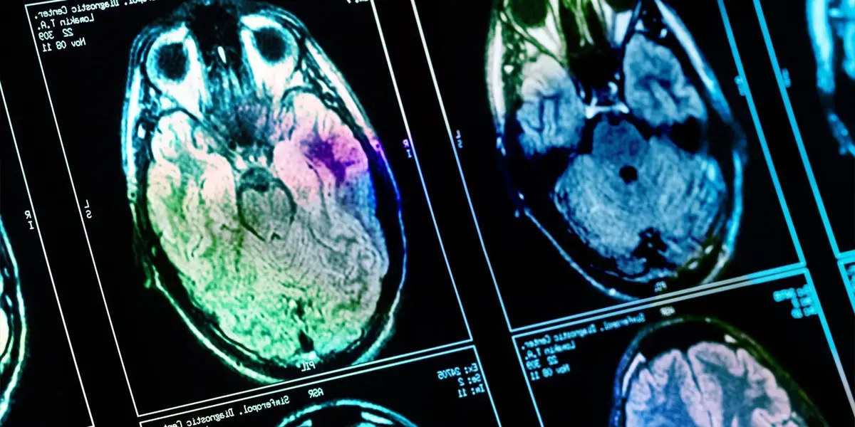
A new study published this month in Biological Psychiatry: Cognitive Neuroscience and Neuroimaging links the microstructure of brain pathways to the intensity of intrusive memories after trauma. Researchers examined survivors of various traumatic events, using advanced MRI techniques to assess white‑matter health and memory symptoms.
Wellness insights
 Brain Wiring Explains Why Trauma Victims Struggle To Find Joy
 Healing Journey: A Therapist Reflects on Processing Deep Childhood Trauma
 Forest walks boost mood through sound and biodiversity awareness
 What Counselling in Two Different Countries Taught Me: Hansel Wong on Connection, Curiosity, and Culture
The team focused on white‑matter tracts that connect emotion‑processing regions with memory centers. Diffusion imaging revealed that reduced integrity in these pathways correlated with more vivid, recurring flashbacks. The findings suggest that physical connectivity, not just psychological factors, shapes how trauma is recalled. Lead author Dr. Elena Martín explained that the results highlight a biological substrate for memory intrusion, offering a potential target for future interventions.
Analysis identified the uncinate fasciculus and the cingulum bundle as critical conduits. Participants with lower fractional anisotropy in these tracts reported higher intrusion scores on standardized questionnaires. The uncinate fasciculus links the amygdala to the prefrontal cortex, regions essential for emotional regulation. Damage or degeneration in this route may impair the brain’s ability to dampen distressing recollections. Similarly, the cingulum connects the hippocampus with broader cortical networks, influencing contextual memory processing. Researchers noted that subtle microstructural changes, invisible to conventional scans, were enough to alter subjective trauma experiences.
Why Do Some People Relive Trauma More Vividly?
The study proposes that individual differences in white‑matter health account for variability in flashback intensity. Genetic factors, chronic stress, and prior injuries can affect myelination, the insulating layer around nerve fibers. When myelin is compromised, signal transmission slows, leading to fragmented yet persistent memory fragments. Dr. Martín added that these neural bottlenecks may prevent the brain from integrating traumatic events into a coherent narrative, leaving them stuck as intrusive images. Understanding this mechanism could inform personalized therapies that aim to strengthen or protect vulnerable pathways.
The implications extend beyond academic insight. Clinicians may soon use diffusion imaging as a diagnostic tool to identify patients at risk for severe intrusive symptoms. Early detection could guide targeted cognitive‑behavioral strategies or novel pharmacological approaches aimed at white‑matter repair. As research progresses, the hope is to transform trauma care from symptom management to underlying circuit restoration.
Frequently Asked Questions
What brain structures were examined? The study focused on the uncinate fasciculus and the cingulum bundle, two white‑matter tracts linking emotion and memory centers.
Can the findings predict who will develop PTSD? While the results suggest a link, they are not yet sufficient for definitive PTSD prediction; further validation is required.
Will this lead to new treatments? Potentially, as identifying vulnerable pathways opens avenues for therapies that protect or restore white‑matter integrity.