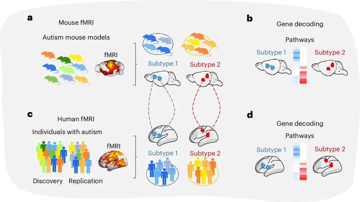
Divergent Connectivity Maps Define Two Autism Profiles
An international team of neuroscientists announced Tuesday that brain scans have identified two separate autism subtypes, each tied to a unique pattern of neural connectivity. The research, coordinated by scientists at Italy’s Institute of Technology, examined functional MRI data from more than 600 participants in Europe and North America between 2022 and 2024.
Wellness insights
 Brain Wiring Explains Why Trauma Victims Struggle To Find Joy
 Healing Journey: A Therapist Reflects on Processing Deep Childhood Trauma
 Forest walks boost mood through sound and biodiversity awareness
 What Counselling in Two Different Countries Taught Me: Hansel Wong on Connection, Curiosity, and Culture
The investigators applied high‑resolution functional MRI to map whole‑brain activity in individuals diagnosed with autism spectrum disorder (ASD). By comparing these maps with those from genetically engineered mouse models, they uncovered consistent dysconnectivity signatures that cluster the human cohort into two groups. The results challenge the notion of a single neurological cause for ASD and suggest that distinct biological pathways underlie the disorder’s diverse symptoms.
One subgroup displayed unusually strong connections between frontal and temporal regions, a pattern linked to heightened sensory perception and repetitive behaviors. „These hyper‑connected circuits may explain why some autistic people experience intense sensory overload,” said Dr. Elena Rossi, the study’s lead author. The second subgroup exhibited weakened links within the default‑mode network, a system crucial for social cognition and self‑reflection. Researchers observed that participants in this group tended to have more pronounced difficulties with social interaction and theory of mind tasks.
Could These Subtypes Guide Personalized Therapies?
Cross‑species analysis reinforced the findings: mice engineered to mimic each connectivity pattern reproduced comparable behavioral traits, confirming that the brain‑wide signatures are not artifacts of human imaging alone. The dual‑type model aligns with earlier genetic studies that hinted at multiple molecular routes to autism, but this is the first work to map those routes onto concrete brain networks.
If autism can be parsed into biologically distinct categories, clinicians may soon tailor interventions to each profile’s neural architecture. „Targeted neurofeedback or pharmacological strategies could be calibrated to either dampen excessive connectivity or boost under‑active networks,” suggested Dr. Marco Bianchi, a co‑author and neuropsychiatrist. The team proposes that future clinical trials stratify participants by subtype, potentially increasing the success rate of experimental treatments that have previously shown mixed results.
The authors caution that larger, longitudinal studies are needed to confirm the stability of these subtypes over time and across developmental stages. Nonetheless, the discovery opens a pathway toward a more nuanced diagnostic framework that moves beyond behavioral checklists to incorporate brain‑based biomarkers.
Frequently Asked Questions
What defines the two autism subtypes identified in the study? The subtypes are distinguished by opposite patterns of brain connectivity: one shows hyper‑connectivity in fronto‑temporal circuits, the other shows hypo‑connectivity in the default‑mode network.
How might these findings affect autism diagnosis? If validated, clinicians could use functional MRI to assign patients to a subtype, enabling more precise prognoses and individualized treatment plans.
Are there immediate therapeutic implications? Not yet. The research suggests possible avenues for personalized interventions, but clinical applications will require further testing and regulatory approval.